September 22, 2025

Ventricular Standstill: Clinical Documentation and Coding Considerations


What is ventricular standstill? 

  • SA node is functioning, and P waves are present on EKG. 
  • There is no ventricular response, no contractions of the muscle. 
  • The presence of complete heart block with no escape rhythm. 
  • No cardiac output with the patient in full arrest. 
  • May be paroxysmal or prolonged. 


Why Ventricular Standstill Is Clinically Significant


  • It is a potentially fatal arrhythmia. 
  • The ventricles come to a complete standstill. 
  • Sustained for more than a few seconds, unconsciousness and no palpable pulse. 
  • Sustained is considered an unshockable rhythm, 10X more deadly than V-fib. 
  • Immediate CPR required with likely placement of a pacemaker. 


Clinical Documentation Tips for Ventricular Standstill

1.) Document the Underlying Cause 


  • Review and query if needed for contributing factors such as: 
  • Electrolyte imbalances (e.g., hyperkalemia, hypokalemia) 
  • Drug toxicity (e.g., cocaine overdose) 
  • Cardiac conditions (e.g., advanced AV block) 
  • Non-cardiac causes (e.g., hypoxia, acidosis) 


2.) Clinical Indicators 


  • Support the diagnosis: 
  • ECG findings (e.g., isolated P waves, absence of QRS) 
  • Symptoms (e.g., syncope, loss of consciousness, pulselessness) 
  • Treatment response (e.g., CPR, pacemaker insertion) 


3.) Specify the Type of Block 


  • If applicable, clarify whether the patient has: 
  • Complete AV block 
  • Trifascicular block 
  • Sub-Hisian block (more life-threatening) 


4.) Treatment Documentation 


  • Note any interventions: 
  • CPR 
  • Temporary or permanent pacemaker 
  • Medications (e.g., epinephrine) 


5.) Avoid Ambiguity 


  • Review for precise terminology. Avoid vague phrases like “heart stopped” or “no rhythm” without correlating ECG or clinical context


Coding Considerations 

1.) Primary Code 


  • I46.9 – Cardiac arrest, unspecified (Classifies as MCC when patient is discharged alive) 


2.) ICD-10-CM Alphabetic Index   

ECG strip illustrating ventricular standstill with absence of ventricular contractions.

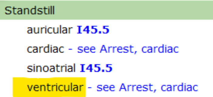
3.) Current Coding Guidance: 

  • When complete/3rd degree heart block is present with asystole in the instance of a brief pause of electrical conduction with spontaneous recovery of sinus rhythm, the asystole is likely caused by the complete heart block. Therefore, only I44.2 is assigned and not reported with the code for cardiac arrest.
  • What does this specifically mean for CDI and Coding? It means we can't code third degree/complete heart block and ventricular standstill together. 


Documentation Example: 

Received page for prolonged AV block. Tele strip shows p waves with ventricular standstill for 19 seconds. Patient is asymptomatic with normal BP at time of arrhythmia. Received IV metoprolol 2.5 mg and now scheduled Q6H for PAF. No other nodal blocking agents. Electrolytes are unremarkable today. TTE was normal this admission. 


Alyce Reavis, RN, MSN, CCDS, CCS, Senior CDI Educator, Consulting Services at UASI

Alyce Reavis, RN, MSN, CCDS, CCS 

Senior CDI Educator, Consulting Services at UASI


Drawing on clinical experience in adult, pediatric, and neonatal acute care, Alyce brings valuable insight to CDI education and documentation improvement. She holds an MSN in Leadership/Education along with CCDS, CCS, and AHIMA’s outpatient CDI micro credential, supporting health systems in strengthening documentation accuracy, quality reporting, and reimbursement integrity. Passionate about truthful, clinically aligned health records, she helps organizations ensure documentation reflects true patient acuity. She is a past presenter for the ACDIS National Convention, Local chapter meetings, and the ACDIS Virtual Best Practices conference. 


Works Cited

Association of Clinical Documentation Integrity Specialists & American Health Information Management Association. (2022). Guidelines for achieving a compliant query practice.


American Hospital Association. (2019). Complete heart block and asystole. Coding Clinic for ICD-10-CM/PCS, Second Quarter, 4.


Centers for Medicare & Medicaid Services. (2025). ICD-10-CM Official Guidelines for Coding and Reporting.
Available at
https://www.cms.gov/medicare/coding/icd10


ICD-10-CM Code Book. (2025).


Prutkin, J. (2024). ECG tutorial: Miscellaneous diagnoses. UpToDate.
Available at
https://www.uptodate.com/contents/ecg-tutorial-miscellaneous-diagnoses


University of New Mexico. (n.d.). Ventricular standstill.
Available at
https://www.ekgstripsearch.com/Vent_SS.htm

MRI brain scans illustrating symptoms of PRES with title text overlay.
December 9, 2025
Learn the symptoms of PRES, key treatment considerations, ICD-10-CM code I67.83, and documentation tips for CDI and accurate DRG assignment.
Microscopic immune cells interacting in cellular environment, illustrating immune effector activity
By Katie Curry December 1, 2025
Understand ICANS documentation and ICD-10 coding with guidance on the ICANS grading system, ICE score, clinical indicators, and CAR T-cell neurotoxicity.
Fingerstick blood glucose test being performed, illustrating screening and monitoring practices for gestational diabetes.
By Katie Curry November 3, 2025
Gestational Diabetes
Clinician pointing to anatomical kidney model illustrating acute kidney injury.
By Katie Curry October 20, 2025
Learn how to identify, document, and code acute kidney injury (AKI), including diagnostic criteria, staging, ICD-10-CM guidance, and CDI query considerations.
Blurred hospital scene symbolizing CDI review of firearm injury intent reporting.
By Katie Curry September 30, 2025
Learn how firearm injury intent is documented and reported in ICD-10-CM, including intent categories, external cause codes, and documentation considerations.
Title image for neurostorming (PSH) documentation and coding
By Katie Curry September 7, 2025
Learn how neurostorming, also known as paroxysmal sympathetic hyperactivity (PSH), is documented and coded using ICD-10-CM guidance.
Title photo of doctor for New ICD-10-CM code E11.A for type 2 diabetes mellitus in remission
By Katie Curry August 7, 2025
FY 2026 ICD-10-CM coding guidance for new code E11.A, Type 2 diabetes mellitus without complications in remission, including documentation and query considerations.
By Katie Curry July 9, 2025
Definition: Neonatal encephalopathy (NE) is a clinically defined syndrome of disturbed neurologic function in the earliest days of life in a term or late preterm infant, manifested by difficulty with initiating and maintaining respiration, depression of tone and reflexes, subnormal level of consciousness, and often seizures. Clinical presentation: Low APGAR scores and/or weak/absent cry in the delivery room. Hyperalert, irritable, lethargic, obtunded. Decreased spontaneous movements, poor tone, blunted or absent primitive reflexes, seizure activity. Breathing and/or feeding difficulties. Documentation Tips: The CDS should review to identify the underlying etiology . (e.g., hypoxic-ischemic event, infection, metabolic disorder). Review clinical indicators that may indicate associated conditions , such as seizures, abnormal imaging, acidosis, or multi-organ dysfunction. Review the documentation for the timing of onset (e.g., at birth, delayed). Common clinical indicators include low APGAR scores, need for resuscitation, abnormal tone, or altered level of consciousness.  ICD-10-CM Coding: P91.811, Neonatal encephalopathy in diseases classified elsewhere P91.819, Neonatal encephalopathy, unspecified Use when the type or etiology of NE is not documented Query Example: To the Attending Neonatologist: Documentation in the medical record indicates the newborn infant delivered from mother with placental abruption demonstrates seizures, abnormal muscle tone, low APGAR scores, and required resuscitation at birth. Imaging showed evidence of cerebral edema. The diagnosis of “neonatal encephalopathy” was documented in the assessment. Query: Based on the clinical indicators, can you clarify the type and cause of the encephalopathy in this newborn? Please select the most appropriate option below or specify another diagnosis: Neonatal encephalopathy due to Hypoxic-ischemic encephalopathy (HIE) Neonatal encephalopathy due to other etiology (please specify) Other (please specify): __________
By Katie Curry May 12, 2025
Definition: Tumor lysis syndrome (TLS) is an oncologic emergency caused by massive tumor cell lysis and the release of large amounts of potassium, phosphate, and uric acid into the systemic circulation. Deposition of uric acid and/or calcium phosphate crystals in the renal tubules can result in acute kidney injury.
Provider with stethoscope, representing clinical evaluation and diagnosis of SIADH and related endocrine fluid imbalances.
By Brandon Losacker April 17, 2025
Syndrome of Inappropriate Antidiuretic Hormone (SIADH) occurs when the pituitary gland releases excessive antidiuretic hormone (ADH) , leading the body to retain fluid and dilute sodium levels in the bloodstream. This condition causes hyponatremia and hypo-osmolality , often triggering a complex clinical picture. What Causes SIADH? SIADH can develop in response to several underlying conditions or external factors: CNS disturbances: Stroke, hemorrhage, infection, and trauma can trigger abnormal ADH release. Cancer: Especially small cell lung cancer, extrapulmonary small cell carcinomas, head and neck cancers, and olfactory neuroblastomas. Medications: SSRIs, NSAIDs, opiates, some antineoplastic drugs, ciprofloxacin, haloperidol, and high-dose imatinib. Surgery: Often linked to pain response. Hormonal deficiencies: Including hypothyroidism and hypopituitarism. Exogenous hormone use: Vasopressin, desmopressin, and oxytocin. HIV infection Hereditary SIADH Diagnostic Criteria: Schwartz and Bartter Clinical Framework A diagnosis of SIADH typically includes: Serum sodium < 135 mEq/L Serum osmolality < 275 mOsm/kg Urine sodium > 40 mEq/L Urine osmolality > 100 mOsm/kg Normal skin turgor and blood pressure (absence of clinical volume depletion) Exclusion of other hyponatremia causes Correction of sodium levels via fluid restriction Important Note: Code only the SIADH, not the hyponatremia, as hyponatremia is considered integral to the disease process . Clinical Scenario A 68-year-old male presents to the ED with confusion , nausea , and a 12-pound weight gain over the past week. He was diagnosed with small cell lung cancer two months ago. Vitals: BP: 160/90 mmHg HR: 110 bpm Labs: Serum sodium: 122 mEq/L Serum osmolality: Decreased Urine: Elevated osmolality and high sodium concentration Indicators Suggestive of SIADH Hyponatremia: Sodium level of 122 mEq/L Diluted Serum Osmolality: From water retention Concentrated Urine: High osmolality and sodium levels despite low serum sodium Recent Weight Gain: 12 lbs in one week, pointing to fluid overload Underlying Malignancy: Small cell lung cancer is a well-known cause of ectopic ADH production Documentation Tips 1. Accurate Diagnosis Clearly state “SIADH” and link it to the underlying cause , such as cancer. 2. Clinical Findings Review provider and nursing notes for symptoms like confusion, nausea, and fluid retention. Confirm vital signs and weight gain. Include lab values: sodium, serum/urine osmolality, and urine sodium. 3. Treatment Plan Document fluid restriction orders . Check MAR for medications such as vasopressin receptor antagonists . Note any improvements in symptoms and lab values after treatment. Tip: High blood glucose can artificially lower serum sodium levels. Use a sodium correction calculator to determine the true sodium level. References Centers for Medicare and Medicaid Services. (2024). ICD-10-CM Official Coding Guidelines. Available here: cms.gov Pinson, R., & Tang, C. (2024). The CDI Pocket Guide. Available here: cdiplus.com Prescott, L., & Manz, J. (2024). ACDIS CDI Pocket Guide. Available here: https://acdis.org/ Sterns, R. (2024). Pathophysiology and etiology of SIADH. UpToDate. Yasir, M., & Mechanic, O.J. (2023). Syndrome of Inappropriate Antidiuretic Hormone Secretion. StatPearls Publishing.
Show More