October 17, 2025
UASI is proud to announce the appointment of Angelica Cage, MBA, BSN, RN, CCDS, CCS, CDIP, CRCR, LSSGB,
as Director of Denials & Strategy.
With a background in critical care nursing, an MBA, and more than a decade of leadership in clinical documentation integrity, coding, denials management, and revenue cycle optimization, Angelica brings a rare blend of clinical expertise and strategic business acumen to the role.
Angelica has successfully led teams and delivered large-scale solutions for some of the nation’s most respected health systems, including Tufts Medicine, and AdventHealth. She has driven significant CDI program enhancements, boosting financial impact by 65% percent within a single year through workflow optimization and cross-functional collaboration.
In addition to her operational leadership, Angelica has earned national recognition for her contributions to the industry. She has been a frequent national speaker for the Association of Clinical Documentation Integrity Specialists, was honored with the ACDIS Professional Achievement Award and the MaHIMA Innovation and Collaboration Award in 2024 and served on the ACDIS Advisory Board. She is an active member of HFMA and AHDAM, contributing to thought leadership and the advancement of best practices in revenue cycle management.
Reflecting on her new role, Cage shared, “Throughout my career, I have been passionate about advancing strategies that bring together the best of clinical expertise and operational performance. UASI is known for its ability to partner with providers in achieving sustainable results, and I look forward to contributing to that legacy by helping clients strengthen compliance, improve documentation and coding integrity, and enhance both financial outcomes and the quality of care delivered to patients.”
Angelica’s appointment underscores UASI’s dedication to bringing forward-thinking leaders onto its team who can bridge clinical insight with operational strategy. By leveraging her expertise, UASI is well positioned to continue delivering innovative mid-revenue cycle solutions that drive compliance, efficiency, and financial performance while supporting the delivery of high-quality patient care.
About UASI
For over 40 years, UASI has bridged the gap between people and processes in financial and clinical operations by providing solutions that reduce revenue leakage, enhance operational efficiency, and ensure quality and compliance.

Clarify apnea of prematurity clinical indicators, treatment considerations, and CDI coding guidance to support accurate neonatal documentation and query decisions.
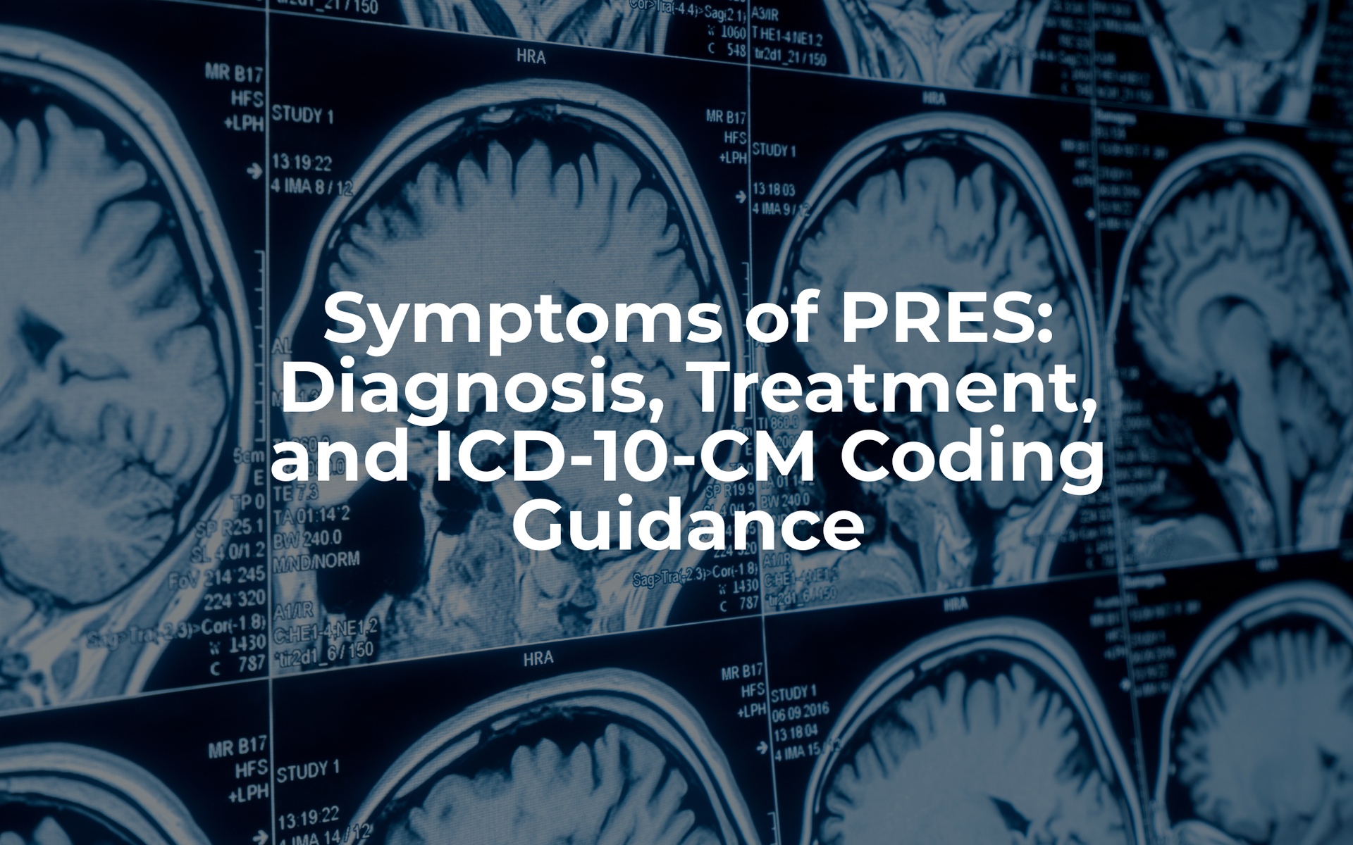
Learn the symptoms of PRES, key treatment considerations, ICD-10-CM code I67.83, and documentation tips for CDI and accurate DRG assignment.

Understand ICANS documentation and ICD-10 coding with guidance on the ICANS grading system, ICE score, clinical indicators, and CAR T-cell neurotoxicity.

Define gestational diabetes documentation requirements, key risk factors, and clinical details needed to support accurate ICD-10 coding, severity capture, and quality reporting.

Learn how to identify, document, and code acute kidney injury (AKI), including diagnostic criteria, staging, ICD-10-CM guidance, and CDI query considerations.
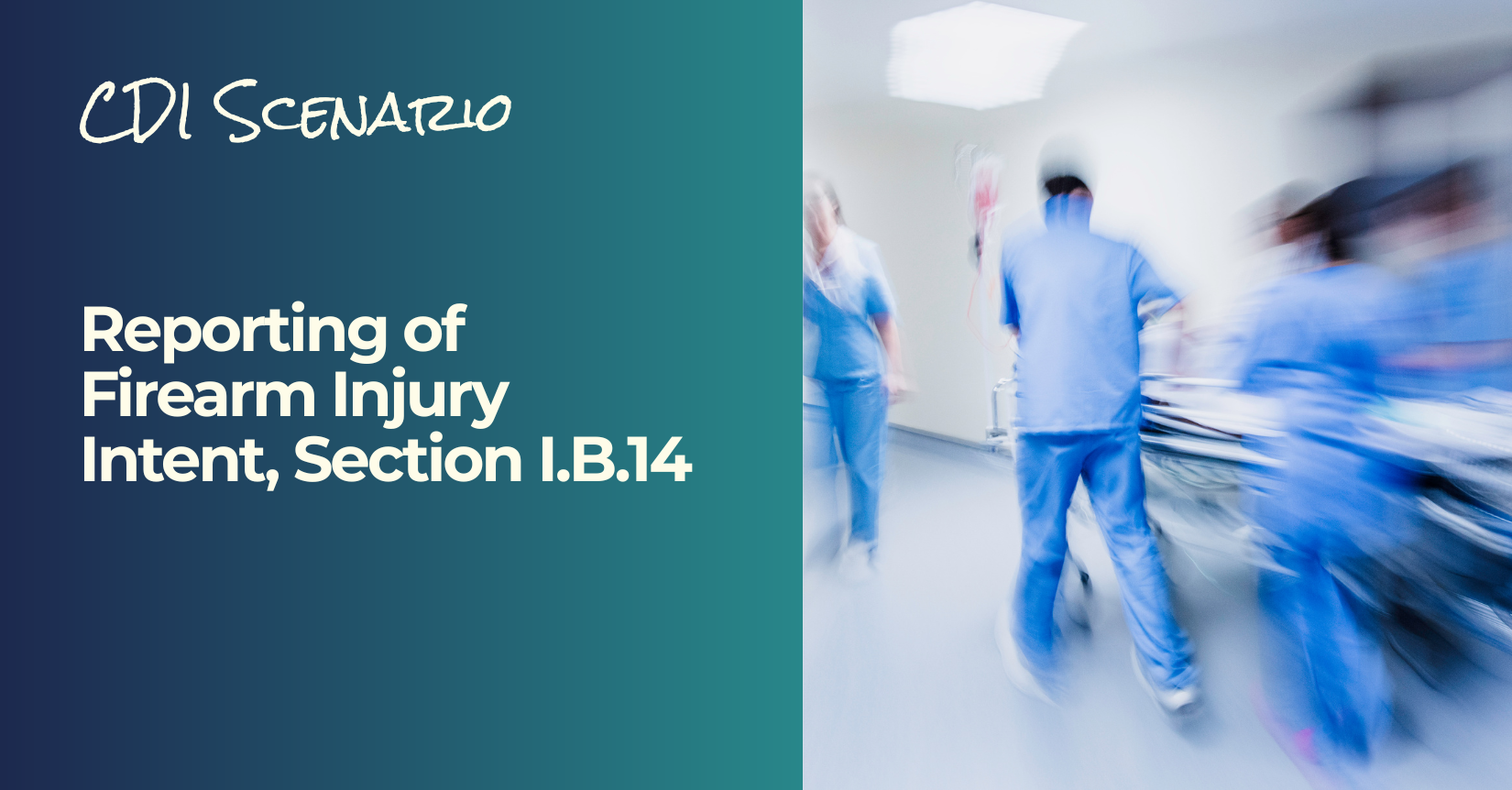
Learn how firearm injury intent is documented and reported in ICD-10-CM, including intent categories, external cause codes, and documentation considerations.
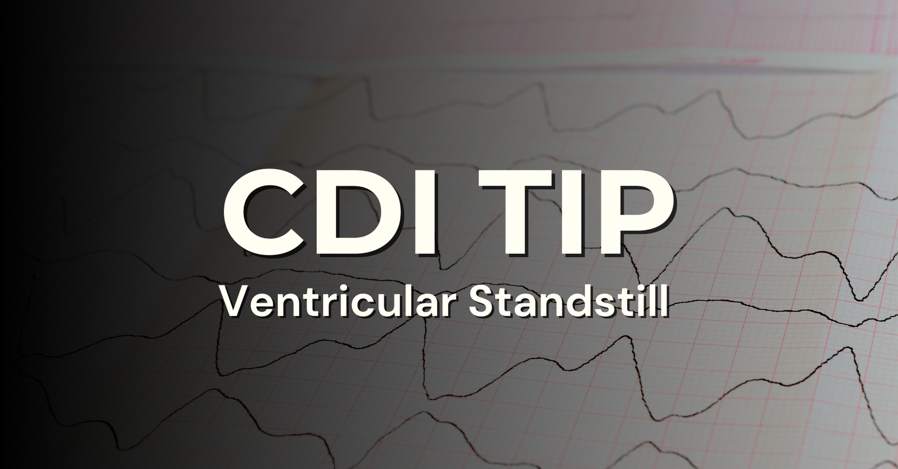
Learn how ventricular standstill is documented and coded, including clinical indicators, ICD-10-CM guidance, and common documentation considerations.

Learn how neurostorming, also known as paroxysmal sympathetic hyperactivity (PSH), is documented and coded using ICD-10-CM guidance.
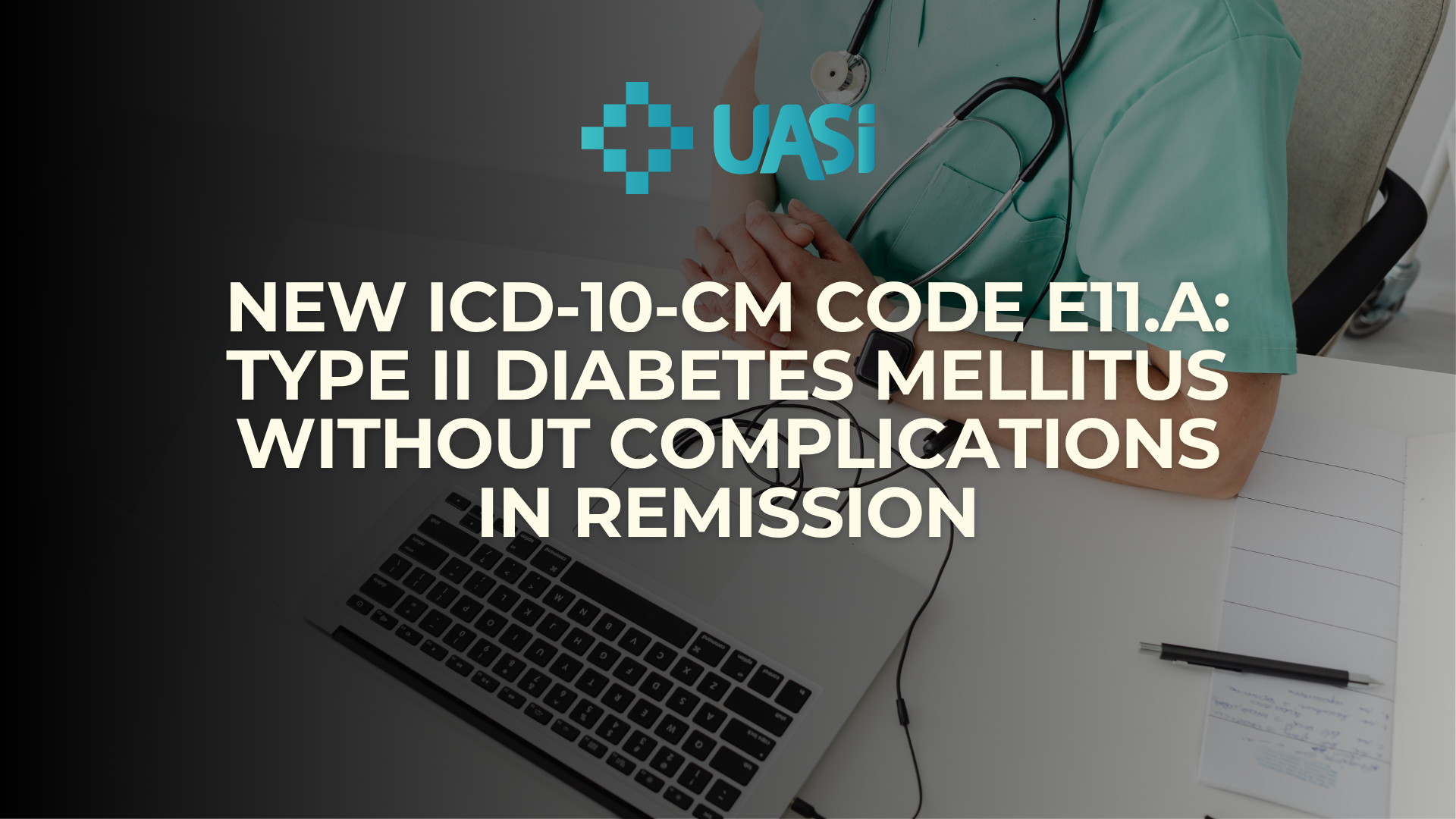
FY 2026 ICD-10-CM coding guidance for new code E11.A, Type 2 diabetes mellitus without complications in remission, including documentation and query considerations.

Clinical documentation and coding guidance for neonatal encephalopathy, including key indicators, query considerations, and ICD-10-CM codes.


